How SCOPE Reagents Are Engineered
The SCOPE Technology Platform
Engineered antigen-based detection reagents for precise, low-background, and reproducible CAR-T flow cytometry.
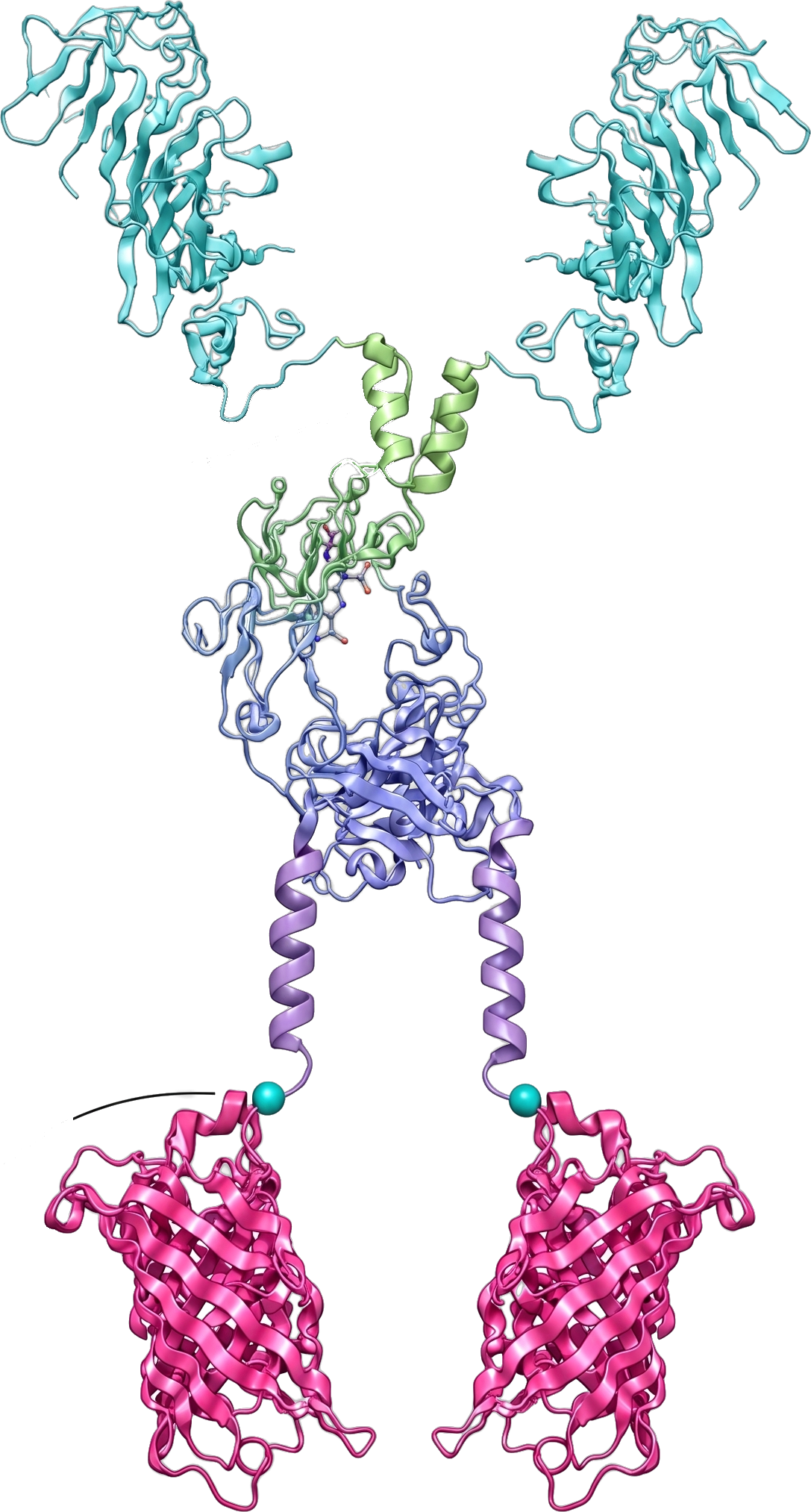
BlueRay SCOPE reagents are designed from the molecular level upward — combining structure-guided antigen engineering, Fc-background reduction, site-specific labeling, and validation on real CAR-T cells to support reproducible CAR-positive cell detection and optional receptor-density analysis.
F:P Ratio
Consistent across lots
Step 01
Optimized Antigen Domain
Structure-guided antigen-domain design preserves CAR-recognition surfaces while improving accessibility, stability, and flow cytometry performance.
Step 02
Engineered Spacer Architecture
Spacer and hinge architecture are optimized to support efficient CAR engagement across different CAR constructs, hinge lengths, and receptor presentations.
Step 03
Reconfigured Fc Region
Fc-region engineering is designed to reduce nonspecific Fc-mediated background and improve separation between CAR-positive and CAR-negative populations.
Step 04
Site-Specific Fluorochrome Conjugation
Site-specific enzymatic labeling supports a defined conjugation architecture, controlled fluorochrome configuration, and batch-to-batch consistency.
Step 05
Defined F:P Ratio
Controlled fluorochrome-to-protein configuration helps reduce lot-dependent variation and supports comparable staining intensity across production batches.
Validated on Real CAR-T Cell Samples
SCOPE reagents are evaluated on real CAR-T cell samples across different CAR constructs, receptor densities, hinge designs, and spacer architectures. This practical validation helps ensure that reagent performance reflects real flow cytometry use cases.
2,000+
CAR-T Cell Samples Analyzed
Supporting robust assay development, reagent optimization, and analytical validation.
Different CAR constructs
Different hinge and spacer formats
Different CAR expression levels
CAR-positive percentage measurement
Optional receptor-density analysis
From Molecular Design to Quantitative Readout
- 1
3D Antigen Design
- 2
Spacer Engineering
- 3
Fc Background Reduction
- 4
Site-Specific Biotinylation
- 5
F:P Control
- 6
Real CAR‑T Validation
- 7
Quantitative Flow Cytometry Readout
- 1
3D Antigen Design
- 2
Spacer Engineering
- 3
Fc Background Reduction
- 4
Site-Specific Biotinylation
- 5
F:P Control
- 6
Real CAR‑T Validation
- 7
Quantitative Flow Cytometry Readout
Engineered for Reproducibility, Not Just Detection
High-Specificity Binding
Structure-guided antigen design for CAR-specific detection.
Very Low Background
Fc-engineered format designed to reduce nonspecific staining.
Batch-to-Batch Consistency
Site-specific labeling and controlled F:P ratio support reproducible performance.
Quantitative Readout Support
Compatible with CAR-positive percentage measurement and optional ACM-based receptor-density analysis.
Research Use Only (RUO): This product is intended for research use only and is not for use in diagnostic or therapeutic procedures.